


Summary
Keratoconus is a condition in which the clear front surface of the eye, the cornea, gradually thins and bulges into a cone shape. It often begins in the teens or twenties, which makes it very different from the age-related conditions most people associate with low vision.
World Keratoconus Day on November 10 is a good moment to talk about it. This guide explains what keratoconus is, how it is treated, and what helps when vision stays limited even after treatment.
What Keratoconus Is
The cornea is normally dome-shaped and smooth, and it does much of the eye’s focusing.
In keratoconus, the cornea weakens and thins, and the pressure inside the eye pushes it outward into a cone.
That uneven shape scatters and distorts light, so the image that reaches the retina is blurred and warped in a way ordinary glasses often cannot fully fix.
Keratoconus usually appears in the teens or early adulthood and tends to progress over years before it levels off. It can affect one eye or both, and often differs between the two. Risk factors include a family history of keratoconus, long-term eye rubbing, and allergic eye disease.
Related reading: Common Genetic Eye Conditions (/common-genetic-eye-conditions/)
Symptoms and How It Progresses
Early keratoconus can be subtle and is easy to mistake for a normal need for stronger glasses. Common symptoms include:
- Blurred or distorted vision that is not fully corrected by glasses
- A prescription that keeps changing, especially increasing astigmatism
- Sensitivity to light and glare, and halos or streaks around lights, especially at night
- Double vision or ghosting in a single eye
- Eye strain, headaches, and trouble reading small print or screens
As keratoconus advances, the cornea can become more irregular, and in some cases scarring develops.
Very rarely, the cornea can swell suddenly, a problem called acute hydrops, which causes sudden pain, redness, and clouded vision and needs urgent care.
How Keratoconus Is Diagnosed
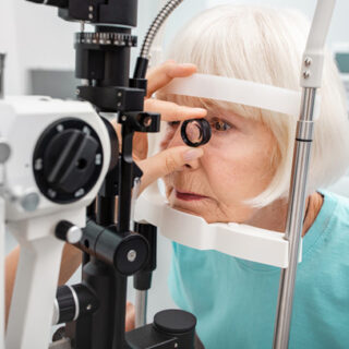
An eye doctor can suspect keratoconus from a routine exam, but confirming it usually involves corneal mapping, called topography or tomography, which measures the shape and thickness of the cornea in detail.
If glasses never seem to fully sharpen your vision, or your prescription keeps changing, ask whether corneal imaging is appropriate.
Treatment Options
Treatment depends on how advanced the condition is and how fast it is changing. An eye care professional will tailor the plan, and most people with keratoconus keep useful vision with modern care. Common options include:
- Glasses or soft contact lenses: helpful in early stages.
- Rigid gas permeable (RGP) lenses: firm lenses that create a smoother optical surface over the irregular cornea.
- Scleral lenses: larger lenses that rest on the white of the eye and vault over the cornea, often comfortable and highly effective for irregular corneas.
- Corneal cross-linking: a procedure that strengthens the cornea to slow or stop progression. It is designed to stabilize the cornea rather than restore lost sharpness.
- Corneal implants or transplant: reserved for more advanced cases where lenses no longer work or scarring limits vision.
Because early treatment can protect vision, it is worth seeing a corneal specialist as soon as keratoconus is suspected.
Avoiding eye rubbing and treating eye allergies are common recommendations too.
When Vision Stays Limited Despite Treatment
For many people, treatment brings vision back to a comfortable level.
For others, even the best lenses, or treatment that arrives after scarring has developed, leave vision that is still blurry, glary, or tiring, particularly for small print, screens, and low-contrast situations.
If that sounds familiar, you are not alone, and it does not mean there is nothing more to do.
A low vision evaluation looks at what you can still see and what tasks are hardest, then matches tools to them. It is a different conversation from a standard eye exam, and it is open to people of any age.
Related reading: Low Vision Eye Diseases (/low-vision-eye-diseases/)
Assistive Technology for Remaining Vision
Assistive technology cannot reshape the cornea, but it can reduce the daily strain that comes with an irregular one. Useful categories include:
- Magnification: handheld, stand, and electronic magnifiers for reading, labels, and detail work.
- Contrast enhancement: boosting the difference between text and background, which can cut through the blur and glare.
- Screen settings: dark mode, larger text, high-contrast themes, and reduced glare on phones and computers.
- Lighting: adjustable task lamps and glare control.
- Wearable vision devices: smart glasses that provide real-time magnification and contrast enhancement for reading, recognizing faces, and using screens.
As with any condition, the right solution depends on how you see and what you want to do, so the best approach is to talk it through with someone who can walk you through the options.
Related reading: Stargardt Disease: Symptoms, Outlook, and Assistive Technology That Helps (/stargardt-disease-symptoms-and-assistive-technology/)
Living and Working with Keratoconus
Keratoconus often arrives when people are in school or building careers, so everyday adjustments matter.
Larger monitors, display settings, good lighting, and regular breaks help. Schools and workplaces often provide accommodations, such as enlarged materials or extra time, once they understand the need.
Take care of your lenses, keep follow-up appointments, and tell your eye doctor right away if your vision suddenly changes.
And connect with others: the National Keratoconus Foundation offers patient resources and community, including its November 10 World Keratoconus Day campaign.
Related reading: Working with Low Vision: Workplace Accommodations and Assistive Technology That Help (/working-with-low-vision-workplace-accommodations/)
Frequently Asked Questions
Is keratoconus a form of low vision?
It can be. Many people with keratoconus see well with treatment, but when vision stays limited even with the best correction, it falls under low vision, and tools designed for low vision can help.
Can keratoconus cause blindness?
Total blindness from keratoconus is rare. Advanced cases can significantly reduce vision, but modern treatments such as specialty lenses, cross-linking, and corneal transplants help most people preserve useful sight.
What causes keratoconus?
The exact cause is not fully understood. Genetics, eye rubbing, and allergic eye disease are all linked to a higher risk.
Can glasses correct keratoconus?
Glasses can help in early stages, but as the cornea becomes more irregular they often cannot sharpen vision fully. Rigid or scleral lenses are commonly used at that point.
Does cross-linking restore lost vision?
Cross-linking is designed to strengthen the cornea and slow or stop progression, not to restore vision that has already been lost. Your eye doctor can explain what to expect in your case.
What to Do Next
If keratoconus has left you with vision that is still hard to work with, it is worth exploring what technology can do. The Vision Buddy team can walk you through how the devices work and whether they fit your vision and daily goals. Reach the Vision Buddy team at hello@visionbuddy.com or (833) 787-2020, or send a message through the contact form on visionbuddy.com.